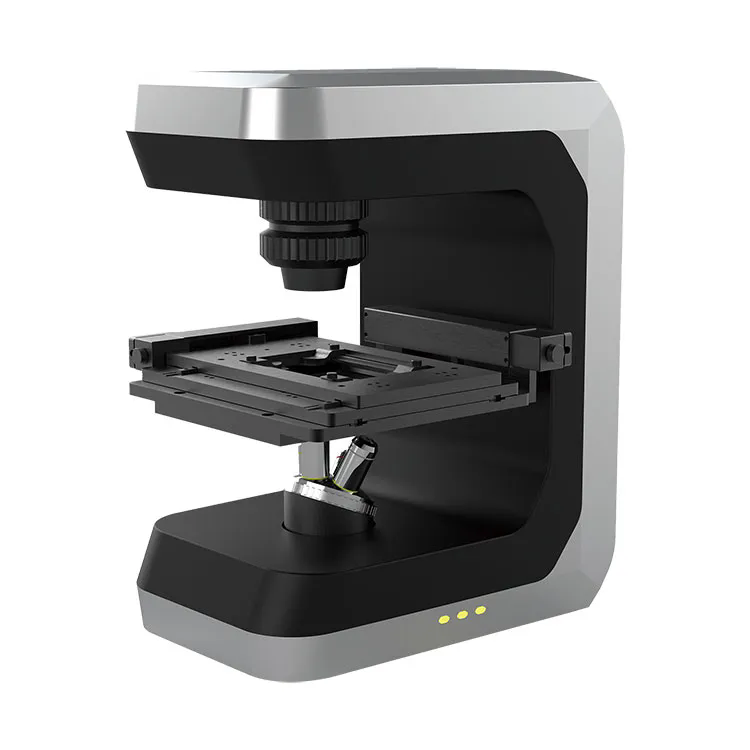
How Does a 3D Cell Microscope Revolutionize Biological Research?
2024-12-06
Microscopy has long been at the heart of scientific discovery, especially in fields like biology and medicine. Traditional microscopes have allowed us to view cells and microorganisms in two dimensions, but what if we could examine biological samples in a more realistic, dynamic way? Enter the 3D cell microscope—a breakthrough technology that’s transforming the way we study cells and tissues. But how exactly does it work, and why is it such a game-changer for research? Let’s explore.
1. What Is a 3D Cell Microscope?
A 3D cell microscope is an advanced imaging system that enables scientists to visualize biological samples in three dimensions. Unlike traditional microscopes, which provide flat, 2D images, 3D microscopes capture detailed, multi-layered views of cells, tissues, and organisms, allowing for a much more comprehensive understanding of biological processes. These microscopes typically utilize techniques such as confocal microscopy, multiphoton microscopy, or light-sheet fluorescence microscopy to create high-resolution 3D images.
2. How Does It Work?
The key to a 3D cell microscope is its ability to gather data from multiple focal planes within a sample, creating a composite image that reveals depth and structure. Here's a basic breakdown of how it works:
- Light Source: A focused laser or light beam is used to illuminate the sample, often with the aid of fluorescent markers.
- Multiple Views: The system captures images from different angles or depths within the sample.
- Data Processing: Advanced software reconstructs these 2D slices into a complete, 3D image, providing a more accurate representation of the sample’s structure.
3. What Makes It Different from Traditional Microscopes?
Traditional microscopes, such as optical and electron microscopes, are limited by their inability to capture the depth and full structure of a sample. They typically offer flat, 2D images, which can make it difficult to fully understand complex biological systems. On the other hand, a 3D microscope allows for:
- Depth Perception: The ability to see through layers of a sample, providing a complete, three-dimensional view.
- Dynamic Imaging: Many 3D microscopes can capture live-cell images, allowing researchers to study biological processes as they unfold in real-time.
- Higher Resolution: 3D microscopes often provide more detailed images, revealing finer cellular structures that might be missed with 2D imaging.
4. How Does It Benefit Biological Research?
The 3D cell microscope is revolutionizing biological and medical research in numerous ways:
- Better Understanding of Cell Function: With 3D imaging, researchers can observe cells in their natural environment, improving our understanding of their functions, interactions, and behaviors.
- Advanced Tissue Studies: Researchers can explore complex tissues and organs in 3D, which is especially useful for studying diseases like cancer, where cell behavior and tissue architecture play crucial roles.
- Live-Cell Imaging: The ability to capture live, dynamic cellular processes, such as cell division, protein trafficking, and signal transduction, offers unprecedented insights into biological mechanisms.
- Improved Drug Development: 3D cell imaging can aid in drug discovery by allowing scientists to observe how new compounds affect cells in a more realistic setting.
5. Key Applications in Medicine and Research
3D cell microscopes have vast applications in various research and medical fields, including:
- Cancer Research: They help study tumor progression, metastasis, and the effects of therapies on cancer cells in three dimensions.
- Neuroscience: Researchers can examine neural networks and brain tissue to gain insights into brain function and neurological diseases.
- Regenerative Medicine: 3D imaging aids in the study of stem cells and tissue engineering, advancing our understanding of tissue regeneration and repair.
- Microbiology and Infectious Diseases: 3D microscopes allow for a more detailed observation of pathogens and their interactions with host cells, improving diagnosis and treatment strategies.
6. The Future of 3D Microscopy
The future of 3D cell microscopy holds exciting possibilities. With continued advancements in technology, 3D microscopes are becoming more accessible and capable of even higher resolutions, making them essential tools in scientific labs around the world. Key areas of development include:
- Faster Imaging: As systems become more efficient, 3D microscopes will be able to capture more data in less time, opening the door for faster research and clinical applications.
- Increased Integration with AI: Artificial intelligence and machine learning can assist in analyzing complex 3D datasets, making it easier to extract meaningful insights from vast amounts of imaging data.
- Non-Invasive Imaging: New technologies may allow for even more detailed 3D imaging of live organisms without damaging or altering the sample, enhancing research in developmental biology and real-time monitoring of disease progression.
7. How Can Researchers Make the Most of 3D Cell Microscopes?
To fully leverage the potential of 3D microscopes, researchers should:
- Get Trained: Proper training in handling and analyzing 3D data is essential to maximize the benefits of these advanced systems.
- Use High-Quality Samples: Proper sample preparation ensures accurate and clear imaging, which is crucial for obtaining high-quality 3D images.
- Collaborate Across Disciplines: The combination of expertise from biologists, chemists, engineers, and data scientists can lead to more innovative applications of 3D microscopy in research and clinical settings.
Why Is a 3D Cell Microscope a Game-Changer?
The 3D cell microscope is not just an evolution of traditional microscopy; it’s a revolutionary tool that is expanding the possibilities of biological research. By providing a deeper, more comprehensive understanding of cellular structures and behaviors, this technology is paving the way for breakthroughs in medicine, drug development, and our fundamental understanding of life itself. As these microscopes continue to improve, they will undoubtedly play an increasingly important role in the future of scientific discovery.
With its ability to capture the complexity of life in three dimensions, the 3D cell microscope has become an indispensable tool in biological and medical research, offering insights that were once unimaginable.


